臨床画像
- EagleView ワイヤレスプローブ型超音波スキャナーで記録
臨床画像
- EagleViewポケットワイヤレスプローブ型超音波スキャナーで記録

心臓
Hypertensive Cardiomyopathy
Hypertensive Cardiomyopathy
Aortic Arch Branches with Color Mode
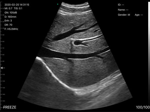
肝臓

肝臓の左葉
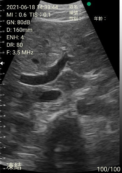
ポータルエリア

肝臓の右葉

肝腎窩

総胆管
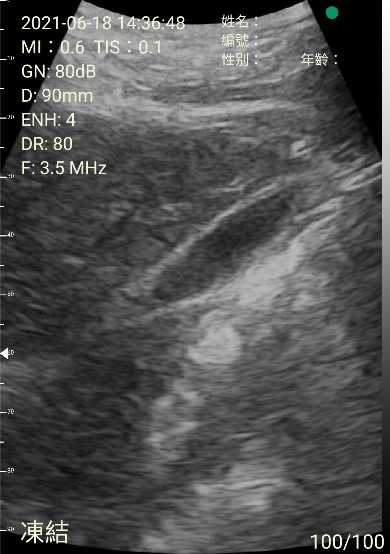
胆嚢
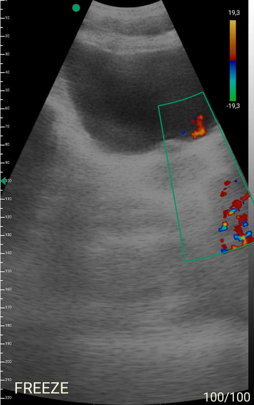
フルブラダー
肺
B lines suggestive of the alveolar – interstitial syndrome

胸水

Renal stone within the right kidney
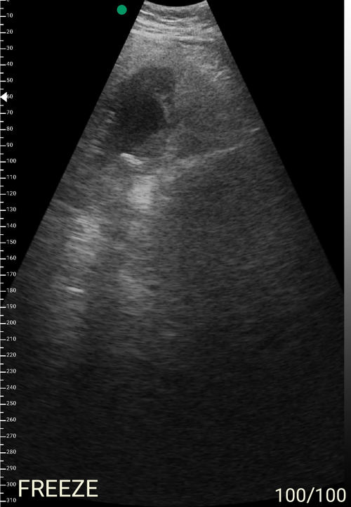
胆嚢
A gallstone in a contracted gallbladder with an associated acoustic shadow.

腹部
Demonstration of bilateral pleural effusion. The picture shows a small amount of pleural effusion with a collapsed lung, diaphragm, and spleen.

腹部
A large amount of pleural effusion, diaphragm and liver

肺
心臓
Big mitral valve vegetations
心臓
Big mitral valve vegetations
心臓
Lung ultrasound in the critically ill (LUCI)
Lung ultrasound in the critically ill (LUCI)

頚動脈

手首の容器

Right common carotid artery with color dopplered

頸静脈血栓症
Carotid artery with color doppler (live)
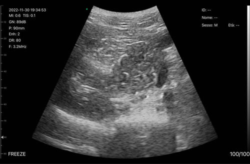
Lliac Arteries

Lliac Arteries

A cortis embolism

A cortis embolism (with pulse wave)

Visualize musculoskeletal pathological conditions

Visualize musculoskeletal pathological conditions
Ultrasound-guided hip injection
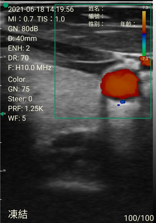
Thyroid and common carotid artery with color doppler

Demonstration of left common carotid artery and internal jugular vein in B mode

Demonstration of left common carotid artery and internal jugular vein with color doppler

Thyroid and common carotid artery in B mode

Thyroid and common carotid artery with color doppler

甲状腺峡部と気管

Thyroid and common carotid artery with color doppler

甲状腺結節

心臓
Hypertensive Cardiomyopathy
Hypertensive Cardiomyopathy
Aortic Arch Branches with Color Mode

肝臓

肝臓の左葉

ポータルエリア

肝臓の右葉

肝腎窩

総胆管

胆嚢

フルブラダー
肺
B lines suggestive of the alveolar – interstitial syndrome

胸水

Renal stone within the right kidney

胆嚢
A gallstone in a contracted gallbladder with an associated acoustic shadow.

腹部
Demonstration of bilateral pleural effusion. The picture shows a small amount of pleural effusion with a collapsed lung, diaphragm, and spleen.

腹部
A large amount of pleural effusion, diaphragm and liver

肺
心臓
Big mitral valve vegetations
心臓
Big mitral valve vegetations
心臓
Lung ultrasound in the critically ill (LUCI)
Lung ultrasound in the critically ill (LUCI)

頚動脈

手首の容器

Right common carotid artery with color dopplered

頸静脈血栓症
Carotid artery with color doppler (live)

Lliac Arteries

Lliac Arteries

A cortis embolism

A cortis embolism (with pulse wave)

Visualize musculoskeletal pathological conditions

Visualize musculoskeletal pathological conditions
Ultrasound-guided hip injection

Thyroid and common carotid artery with color doppler

Demonstration of left common carotid artery and internal jugular vein in B mode

Demonstration of left common carotid artery and internal jugular vein with color doppler

Thyroid and common carotid artery in B mode

Thyroid and common carotid artery with color doppler

甲状腺峡部と気管

Thyroid and common carotid artery with color doppler

甲状腺結節
今すぐEagleViewを入手してください。
XNUMX回限りの購入。 会員登録は必要ありません。
製品を見る超音波ガイド下中心静脈カテーテル留置:構造化されたレビューと臨床診療のための推奨事項
中心静脈カテーテル(CVC)の留置は、集中治療医学および麻酔科での日常的な手順ですが、急性の重篤な合併症(動脈穿刺またはカニューレ挿入、血腫、血胸、または気胸など)は、関連する割合の患者で発生します。 超音波(US)の使用は、CVC合併症の数を減らし、CVC留置の安全性と品質を高めるために提案されています。
続きを読む手順ガイダンスのための超音波
POCUSは、中心静脈カテーテル挿入、困難な末梢動脈および静脈カテーテル挿入、関節穿刺、気道管理、胸腔穿刺、穿刺、腰椎穿刺、局所神経ブロックなどのさまざまなED手順をガイドするために使用されます。 手順のガイダンスにPOCUSを使用すると、成功率が向上し、合併症率が低下し、中心静脈カテーテル挿入の標準治療と見なされます。
続きを読む上肢および下肢ブロックの超音波ガイダンス
超音波ガイダンスのみ、またはPNSと組み合わせて実行される末梢神経ブロックは、感覚および運動ブロックの改善、補給の必要性の減少、および報告された軽度の合併症の減少という点で優れているという証拠があります。 超音波を単独で使用すると、神経刺激と比較してパフォーマンス時間が短縮されますが、PNSと組み合わせて使用すると、パフォーマンス時間が長くなります。
続きを読む肺超音波:心臓専門医のための新しいツール
長年にわたり、肺は超音波検査の立ち入り禁止と見なされてきました。 ただし、最近、肺超音波(LUS)が心血管疾患の多くの肺の状態を評価するための有用なツールとなる可能性があることが示されました。 心臓専門医のためのLUSの主な用途は、Bラインの評価です。 Bラインは、水で厚くなった肺小葉間中隔に由来する残響アーチファクトです。 複数のBラインが肺うっ血に存在し、血管外肺水の検出、半定量化、モニタリング、呼吸困難の鑑別診断、慢性心不全と急性冠症候群の予後の層別化に役立つ可能性があります。
続きを読む救急科におけるポイントオブケア超音波
ポイントオブケア超音波(POCUS)は有用な診断ツールであり、救急科で提供されるケアの不可欠な部分になっています。 過去13年間で、診断および治療スキルを含むように進化してきました。 POCUSは、救急医が診断精度を向上させ、全体的な患者ケアを向上させるのに役立ちます。 この章では、すべての救急医の診断装備内で考慮されるXNUMXのコアPOCUSアプリケーションを要約します。
続きを読むポイントオブケア超音波:プライマリケアにすぐに来る?
これらすべてのアプリケーションで、POCUSは安全で正確、そして有益であり、かかりつけの医師を含む非放射線専門医による比較的少量のトレーニングで実行できます。
続きを読む
モバイルデバイスを使用している場合は、左右の矢印を使用してスライドショーをナビゲートするか、左右にスワイプします


